Veterinarians at the University of Pennsylvania’s veterinary school are studying a type of robotic computed tomography (CT) scan they hope can advance medical care for horses as well as humans. If successful, this technology could eliminate the need for people to lie still inside a claustrophobic tube.
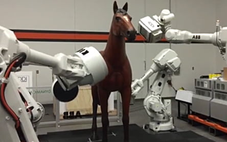
Robotic CT allows a horse to remain awake and standing as scanners on mechanized arms move around it providing high-quality images. The technology even provides some 3D imaging. Additionally, the innovation of this type of CT scan generates anatomical views of the horse in its normal, upright state. This new type of scan is an enormous technical advance compared to current horse CT scans which require administering anesthesia, placing the animal on its side and moving the unit around the effected area. Moreover, all parts of the horse’s body cannot even fit in today’s scanners. The advantage to CT is that unlike X-rays, it is able to provide pictures of soft tissue.
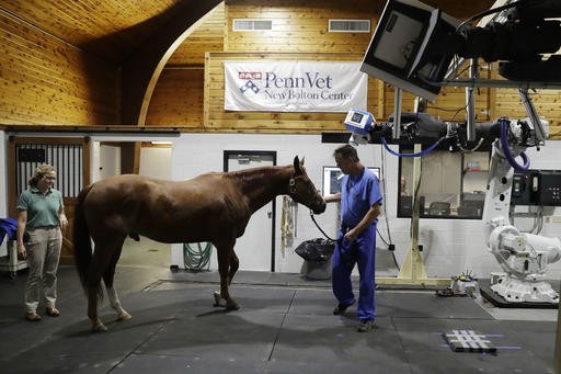
Dr. Barbara Dallap Schaer said, “[Robotic CT] is much less stressful.” Schaer, the medical director of Penn Vet’s New Bolton Center, also points out that, “It’s a pretty athletic event for horses to recover from general anesthesia.” While traditional CT requires the subject to be still, the new system allows for slight movement. At the New Bolton Center, horses are given a mild sedative and walked into the facility for a scan that lasts less than a minute. The hope is that the system’s ability to compensate for movement will eventually advance to the point of being able to capture CT images of a horse running on a treadmill.
New Bolton chief of surgery Dr. Dean Richardson points out that the ease of imaging will enable more horses to receive preventive scans. Many horse owners today are hesitant to allow CT diagnostic procedures due to the potential risk of injury, even catastrophic, caused by the treacherous nature of recovering from anesthesia. “So the whole beauty of this technology, we hope, is that we’re going to be able to scan much greater numbers of patients, much earlier in the process of things like stress-related injuries in a racehorse,” says Richardson.
Penn’s translational research team has partnered with other hospitals to explore utilizing the technology for humans. For people, the science could aid when working with squirming children or claustrophobic adults. Doctors could also get better views of some ailments. For example, a CT scan performed on a patient standing as opposed to lying down could provide a physician a clearer view of a spinal problem. Dr. Raul Uppot is an assistant professor of radiology at Harvard Medical School not involved in the research. He says, “[The technology] does offer something that doesn’t currently exist in the market (for humans).”
Penn was the first to acquire this new system called Equimagine (cost $545,000). The 4DDI company based in New York created the system with components from robot manufacturer ABB. According to 4DDI CEO Yiorgos Papaioannou, the company now has orders for the machine at more than a dozen equine facilities around the world and says, “The word is spreading.” The cost of the new system, while not inexpensive, is comparable to standard CT scanners. The one at Penn was made possible through a donor. 4DDI plans to make a version of the system for smaller animals.
This article was adapted from an AP story by Kathy Matheson titled “Robotic scan for horses could hold promise for human health.”